Angiography in Real-time from 2D to 3D using by AI
Viewing images of the inside of the body in 3D allow for more intuitive manupulation
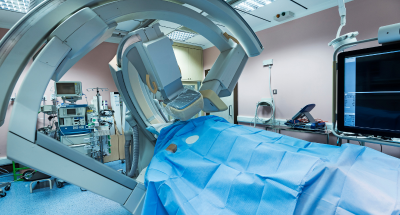
What is the Angiography?
Angiography is an examination in which a tube called a catheter is inserted into the target blood vessel and a contrast medium is administered to depict blood vessels that are not normally visible on simple radiography or X-ray fluoroscopic.Angiography uses contrast medium and carries it into the peripheral organs through the blood flow caused by cardiac output. The angiogram provides a series of images of the circulatory dynamics of the arteries, capillaries, and veins.
Solution & benefits
Viewing images of the inside of the body in 3D allows for more intuitive manipulation. Imagine, for example, looking into the lungs. Let’s start with an endoscope. When looking at the trachea through an endoscope, you can see the trachea beyond the endoscope, but you may not know exactly how much further you need to extend it in the endoscope image. This is because the depth of the trachea cannot be seen clearly in 2D image. By using a 3D monitor to view the endoscopic image, the depth of the endoscope can be clearly seen. This improves the operability of the endoscope. Also,
by showing the images to beginners, it can be useful to improve their learning. Let’s say that the image is viewed with an x-ray.
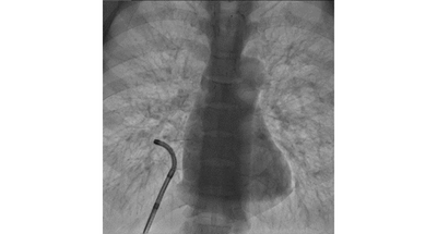
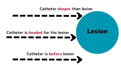
By using a 3D monitor, it is difficult to determine whether the catheter is bent toward the front or the back of the lesion, but by using a 3D monitor, it is possible to get a sense of depth. By using a 3D monitor, you can see where the catheter is in front of, behind, or in the center of the lesion.
Main focus and Depth of Field
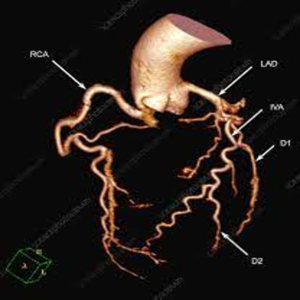
Coronary Angiography
Image on the Under is an ANGIO image of a coronary artery. This image shows contrast examination in real-time, and is the first in the industry to allow 3D imaging from any angle using a biplane angiography.
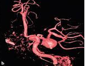
Cerebral angiography
The image can be taken from any angle.This image was taken from the side, and the area that is just in the center, round, and tightly fringed with black color is the cerebral aneurysm.