Notice of Commencement of Multi-institutional Joint Research on quasi-3D fluoroscopic images using AI Technology
This announcement has been officially approved by the public relations of NCC (National Cancer Center Hospital) and Asahi General Hospital
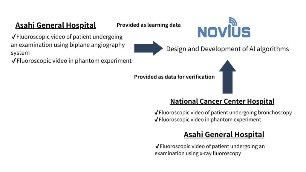
Novius Company, ltd is pleased to announce that it has signed off the multi-institutional joint research agreement with NCC (National Cancer Center Hospital) and Asahi General Hospital, the research started on April 1, 2024.


<Title of Research>
The multi-institutional observational study on quasi-3D of fluoroscopic images using AI technology.
<Research Overview>
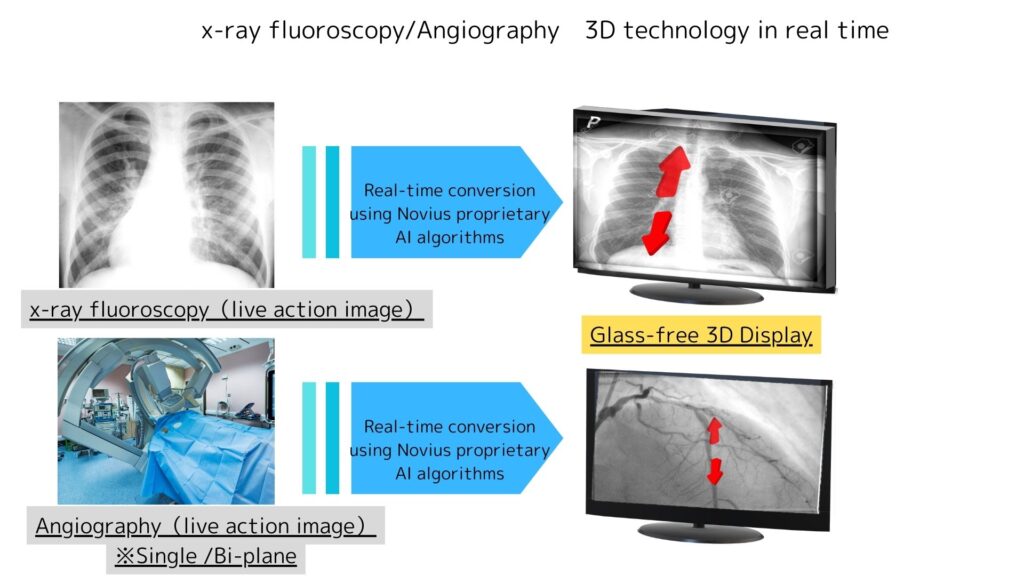
The objective of this research is to enable real-time recognition of the “front” and “back” of flat fluoroscopic image by converting it
to quasi-3D, thereby facilitating guidance to the lesion. We will also continue research and development on the 3Dization of planar images using biplane angiography system at the same time.
<Planned research person who applies>
Patients who underwent bronchoscopy at the National Cancer Center Hospital. Patients who underwent an examination using a biplane angiography device or x-ray fluoroscopy device at the NCC Hospital and Asahi Center Hospital
<Term of Study>
April 1, 2024, to March 31, 2028
<Research Background>
Conventional bronchoscopy biopsy using only x-ray fluoroscopy has an inadequate diagnostic yield because of uncertainty in assessing whether the biopsy instrument has reached the lesion due to difficulty in grasping the three-dimensional positioning of the lesion. The reason for this is that the image depicted by fluoroscopy is two-dimensional, and at least the frontal image alone is not suitable for understanding anterior-posterior misalignment and distance.
<Future Prospects with Technologies to be Researched and Developed>
Real-time 3D fluoroscopic images using AI x 3D conversion technology,
By using AI x 3D conversion technology to convert fluoroscopic images into real-time 3D image
・The location and orientation of catheters in the body can be visualized in more detail and with greater accuracy.
・Real-time 3D images enable physicians to visualize the relationship between the catheter and surrounding tissue in three dimensions.
The real-time 3D image allows physicians to visualize the relationship between the catheter and surrounding tissues in three dimensions,
and to evaluate the catheter appropriately based on these images, thereby improving the accuracy and safety of bronchoscopy.
In cardiovascular procedures, real-time 3D images enhance visualization of complex cardiac anatomy and can guide interventions such as
coronary angioplasty and stent placement.
<Title of Research>
The Multi-institutional observational study on quasi-3D of fluoroscopic images using AI technology
<Research Implementation Structure>
(Leader of Research Project and Principal Researcher)
Yuji Matsumoto MD
Department of Endoscopy (Respiratory Organs),
National Cancer Center Hospital
(Principal Researcher)
Jun Isobe MD, Chief Radiologist & General Manager of Department of Radiology,
Asahi General Hospital
(Principal Researcher)
Koji Okawa, Chief Architect of AI Development
Novius Company,ltd
(Research Administration Office)
Yukihiro Yoshida,MD, Department of Pulmonary Surgery,
National Cancer Center Hospital
Postal code 104-0045
Address: 5-1-1 Tsukiji, Chuo-Ku, Tokyo, Japan
TEL: 03-3542-2511

